In natural environments, microorganisms usually exist as mixed populations. However, if we are to study, characterize, and identify microorganisms, we must have the organisms in the form of a pure culture. A pure culture is one in which all organisms are descendants of the same organism. Techniques for obtaining pure cultures from a mixed population will be described in Lab 3.
In working with microorganisms we must also have a sterile nutrient-containing-medium in which to grow the organisms. Anything in or on which we grow a microorganism is termed a medium. A sterile medium is one which is free of all life forms. It is usually sterilized by heating it to a temperature at which all contaminating microorganisms are destroyed.
Finally, in working with microorganisms, we must have a method of transferring growing organisms (called the inoculum) from a pure culture to a sterile medium without introducing any unwanted outside contaminants. This method of preventing unwanted microorganisms from gaining access is termed aseptic technique.
Keep in mind that you must wear the correct Personal Protection Equipment (PPE) in all labs where you are using microbial cultures, stains, chemicals, and glassware or microscope slides!
A. ASEPTIC TECHNIQUE
Videos reviewing techniques used in this lab:
Forms of Culture Media
Aseptic Technique Tips
Aseptic Technique: Inoculation of broth tubes, slant tubes, and stab tubes
Aseptic Technique: Inoculating a Petri plate - Streaking for Isolation
The procedure for aseptically transferring microorganisms is as follows:
1. Using a bacto-incinerator to sterilize the inoculating loop
Bacto-incinerators enable the sterilization of inoculating loops without having to use an open flame of a Bunsen burner. The bacto-incinerator uses infrared heat at a temperature of 816°C to sterilize the wire portion of the inoculating loop. Be careful not to touch the top portion of the bacto-incinerator. It becomes very hot!
a. At the start of class. turn on the bacto-incinerator and wait 10 minutes for it to heat up.
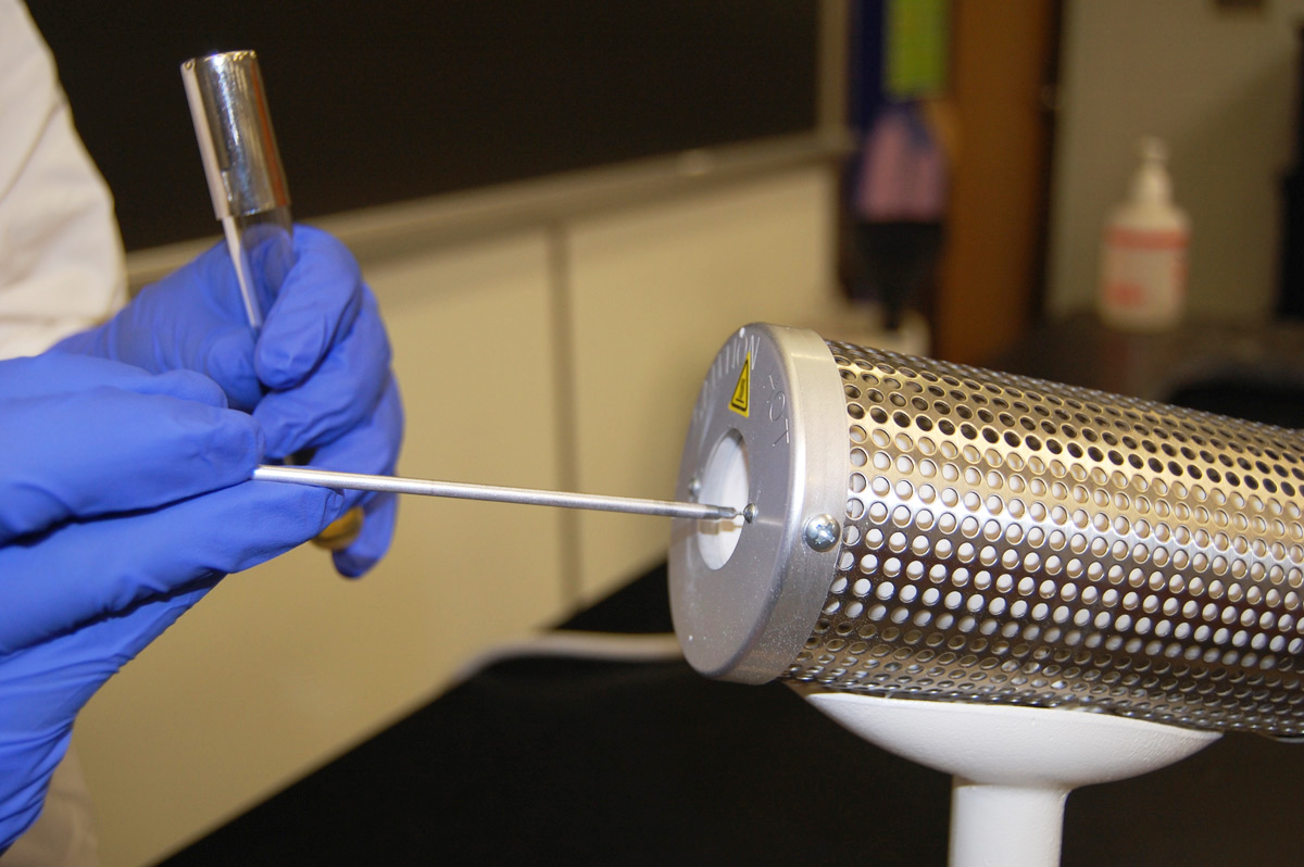
b. Place the entire wire portion of the inoculating loop into the opening of the bacto-incinerator and hold it there for 10 seconds. (See Fig. 1.) In this way all contaminants on the wire are incinerated. To avoid burning your hand on the handle of an overheated inoculating loop, never lay the loop down in the bacto-incinerator and then attempt to pick it up. Never lay the loop down once it is sterilized or it may again become contaminated.
Fig. 1: Using a Bacto-incinerator to Sterilize the Inoculating Loop
Sterilize the inoculating loop by placing it in the bacto-incinerator for 10 seconds.Gary E. Kaiser, Ph.D.
Professor of Microbiology
The Community College of Baltimore County, Catonsville Campus
This work is licensed under a Creative Commons Attribution 3.0 Unported License

c. Allow the loop to cool 10-20 seconds before removing the inoculum.
2. Remove the inoculum.
a. Removing inoculum from a broth culture (organisms growing in a liquid medium):
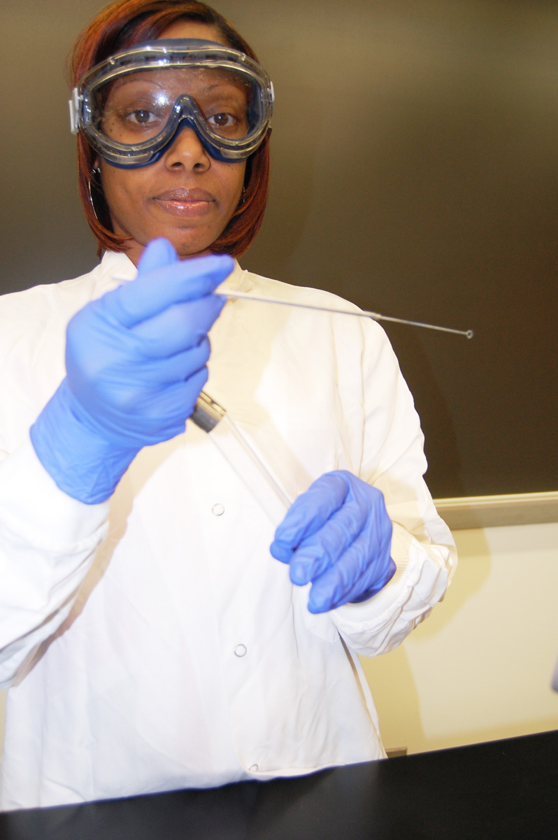
1. Hold the culture tube in one hand and in your other hand, hold the sterilized inoculating loop as if it were a pencil. (See Fig. 1A.)
Fig.1A: Holding the Inoculating Loop and the Culture Tube
Hold the culture tube in one hand and the inoculating loop in the other.Gary E. Kaiser, Ph.D.
Professor of Microbiology
The Community College of Baltimore County, Catonsville Campus
This work is licensed under a Creative Commons Attribution 3.0 Unported License
2. Remove the cap of the pure culture tube with the little finger of your loop hand. (See Fig. 1B and Fig. 1B2.) Never lay the cap down or it may become contaminated.
Fig. 1B: Removing the Cap of the CultureTube
Fig. 1B2: Removing the Cap of the CultureTube
Using the little finger of your loop hand, remove the cap of the pure culture tube.
Wrap your little finger of your loop hand around the cap. Pull off the cap. Do not set it down.Gary E. Kaiser, Ph.D.
Professor of Microbiology
The Community College of Baltimore County, Catonsville Campus
This work is licensed under a Creative Commons Attribution 3.0 Unported License
Gary E. Kaiser, Ph.D.
Professor of Microbiology
The Community College of Baltimore County, Catonsville Campus
This work is licensed under a Creative Commons Attribution 3.0 Unported License
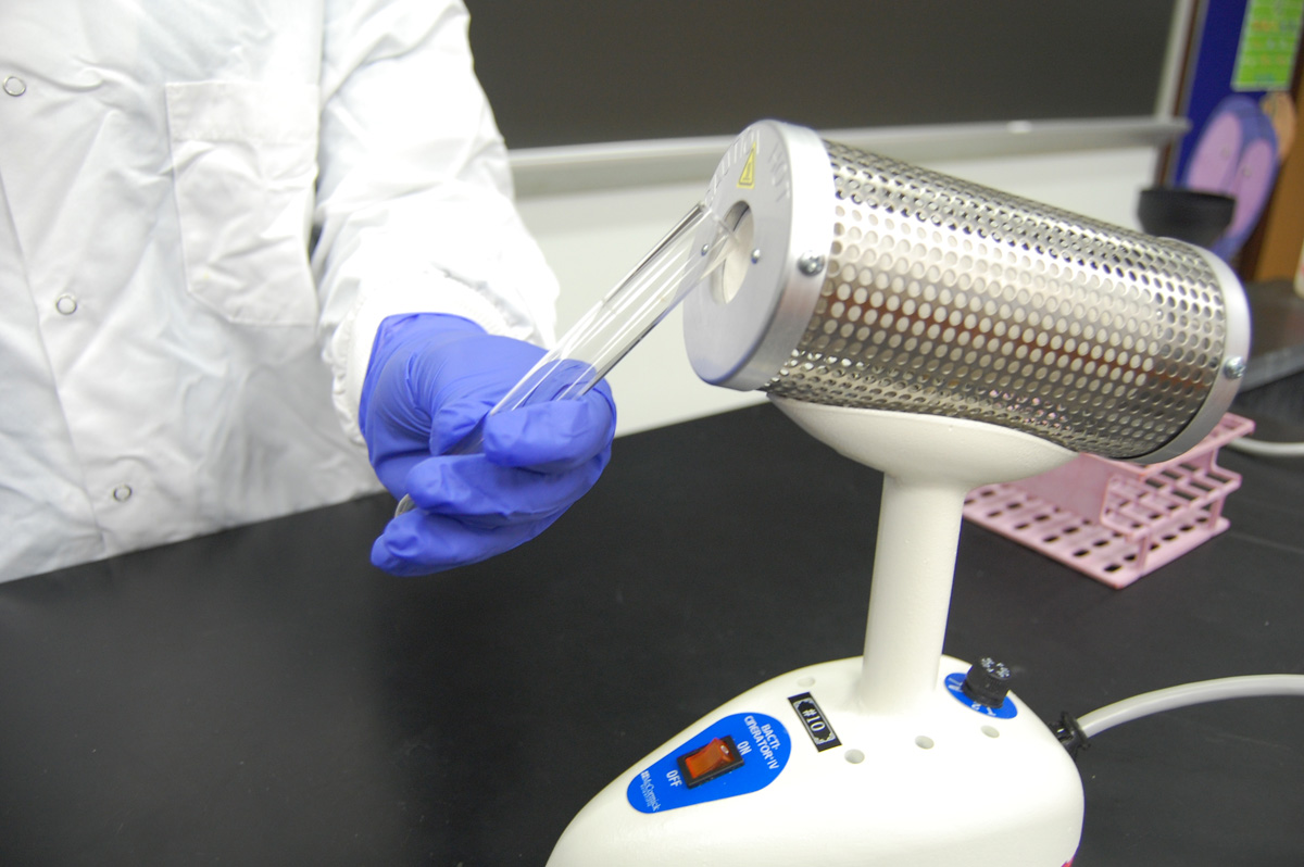
3. Place the lip of the culture tube at the opening of the bacto-incinerator for 2-3 seconds (See Fig. 1C.) This heats the glass and creates a convection current which forces air out of the tube and prevents airborne contaminants from entering the tube.
Fig. 1C: Heating the Lip of the Tube
Place the lip of the culture tube at the opening of the bacto-incinerator for 2-3 seconds.Gary E. Kaiser, Ph.D.
Professor of Microbiology
The Community College of Baltimore County, Catonsville Campus
This work is licensed under a Creative Commons Attribution 3.0 Unported License
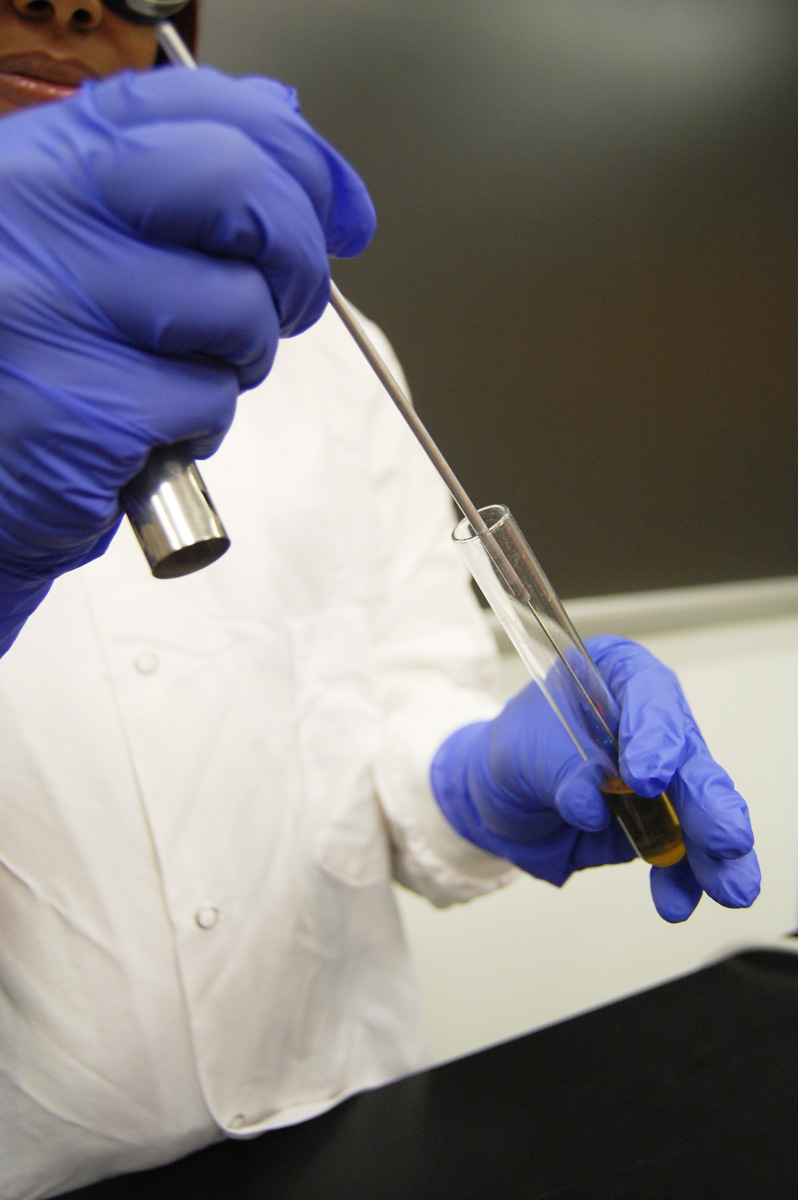
4. Insert the inoculating loop and remove a loopful of inoculum. (See Fig. 1D.)
Fig. 1D: Removing the Bacteria from the Culture Tube
Remove a loopfull of bacteria from your pure culture tube.Gary E. Kaiser, Ph.D.
Professor of Microbiology
The Community College of Baltimore County, Catonsville Campus
This work is licensed under a Creative Commons Attribution 3.0 Unported License
5. Again place the lip of the culture tube at the opening of the bacto-incinerator for 2-3 seconds. (See Fig. 1E.)
Fig. 1E: Heating the Lip of the Tube
Place the lip of the culture tube at the opening of the bacto-incinerator for 2-3 seconds.Gary E. Kaiser, Ph.D.
Professor of Microbiology
The Community College of Baltimore County, Catonsville Campus
This work is licensed under a Creative Commons Attribution 3.0 Unported License
6. Immediately replace the cap. (See Fig. 1F.)
Fig. 1F: Replacing the cap of the Culture Tube
Replace the cap of the pure culture tube.Gary E. Kaiser, Ph.D.
Professor of Microbiology
The Community College of Baltimore County, Catonsville Campus
This work is licensed under a Creative Commons Attribution 3.0 Unported License
Step-by-step summary of removing the inoculum from a broth tube
b. Removing inoculum from a plate culture (organisms growing on an agar surface in a petri plate):
1. Sterilize the inoculating loop by placing it in the bacto-incinerator for 10 seconds. (See Fig. 3A.)
Fig. 3A: Using a Bacto-incinerator to Sterilize the Inoculating Loop
Sterilize the inoculating loop by placing it in the bacto-incinerator for 10 seconds.Gary E. Kaiser, Ph.D.
Professor of Microbiology
The Community College of Baltimore County, Catonsville Campus
This work is licensed under a Creative Commons Attribution 3.0 Unported License
2. Lift the lid of the culture plate slightly and stab the loop into the agar away from any growth to cool the loop.
3. Scrape off a small amount of the organisms and immediately close the lid (See Fig. 3B.)
Fig. 3B: Removing Bacteria from a Petri Plate
Before removing bacteria from the petri plate, first cool the loop by sticking it into the agar away from any growth. Scrape off a small amount of bacteria.Gary E. Kaiser, Ph.D.
Professor of Microbiology
The Community College of Baltimore County, Catonsville Campus
This work is licensed under a Creative Commons Attribution 3.0 Unported License
3. Transfer the Inoculum to the Sterile Medium.
a. Transferring the inoculum into a broth tube:
1. Pick up the sterile broth tube and remove the cap with the little finger of your loop hand. (See Fig. 2A.) Do not set the cap down.
Fig. 2A: Removing the Cap of the Sterile Broth Tube
Using the little finger of your loop hand, remove the cap of the sterile broth tube.Gary E. Kaiser, Ph.D.
Professor of Microbiology
The Community College of Baltimore County, Catonsville Campus
This work is licensed under a Creative Commons Attribution 3.0 Unported License
2. Place the lip of the culture tube at the opening of the bacto-incinerator for 2-3 seconds. (See Fig. 2B.)
Fig. 2B: Heating the Lip of the Tube
Place the lip of the tube at the opening of the bacto-incinerator for 2-3 seconds.Gary E. Kaiser, Ph.D.
Professor of Microbiology
The Community College of Baltimore County, Catonsville Campus
This work is licensed under a Creative Commons Attribution 3.0 Unported License
3. Place the loopful of inoculum into the broth, and withdraw the loop. (See Fig. 2C.) Do not lay the loop down!
Fig. 2C: Inoculating a Sterile Broth Tube with the Bacteria from the Culture Tube
Place bacteria from your pure culture into the sterile broth tube.Gary E. Kaiser, Ph.D.
Professor of Microbiology
The Community College of Baltimore County, Catonsville Campus
This work is licensed under a Creative Commons Attribution 3.0 Unported License
4. Again place the lip of the culture tube at the opening of the bacto-incinerator for 2-3 seconds. (See Fig. 2D.)
Fig. 2D: Heating the Lip of the Tube
Place the lip of the tube at the opening of the bacto-incinerator for 2-3 seconds.Gary E. Kaiser, Ph.D.
Professor of Microbiology
The Community College of Baltimore County, Catonsville Campus
This work is licensed under a Creative Commons Attribution 3.0 Unported License
5. Replace the cap. (See Fig. 2E.)
Fig. 2E: Replacing the cap of the Tube
Replace the cap of the pure culture tube.Gary E. Kaiser, Ph.D.
Professor of Microbiology
The Community College of Baltimore County, Catonsville Campus
This work is licensed under a Creative Commons Attribution 3.0 Unported License
6. Resterilize the inoculating loop by placing it in the bacto-incinerator for 10 seconds. (See Fig. 2F.) Now you may lay the loop down until it is needed again.
Fig. 2F: Using a Bacto-incinerator to Sterilize the Inoculating Loop
Sterilize the inoculating loop by placing it in the bacto-incinerator for 10 seconds.Gary E. Kaiser, Ph.D.
Professor of Microbiology
The Community College of Baltimore County, Catonsville Campus
This work is licensed under a Creative Commons Attribution 3.0 Unported License
Step-by-step summary of transferring the inoculum into a broth tube.
b. Transferring the inoculum into a petri plate:
1. If the agar surface of the plate is visibly wet, use a sterile swab to gently remove the water.
2. On the bottom of the petri plate, divide the plate into thirds with your wax marker and label as shown below. This will guide your streaking.
3. Lift the edge of the lid just enough to insert the loop.
4. Streak the loop across the surface of the agar medium using the either the pattern shown in Figs. 4A-4C or the pattern shown in Fig. 5A-5E.
Fig. 4A: Streaking For Isolation, Step-1
Fig. 4B: Streaking For Isolation, Step-2
Fig. 4C: Streaking For Isolation, Step-3
Streak the inoculum over area "1" as shown above. Then flame the loop and cool it by sticking it into the edge of the agar. Rotate the plate 90 degrees counterclockwise.
Rotate the plate so area "1" is on your left. Drag your stetile inoculating loop through area "1" two times and spread out over area "2" as shown above. Then flame the loop and cool it by sticking it into the edge of the agar. Rotate the plate 90 degrees counterclockwise.
Rotate the plate so that area "2" is on your left. Drag your sterile inoculating loop through area "2" two times and spread the inoculum on area "2" over area "3." Sterilize the loop.Gary E. Kaiser, Ph.D.
Professor of Microbiology
The Community College of Baltimore County, Catonsville Campus
This work is licensed under a Creative Commons Attribution 3.0 Unported License
Gary E. Kaiser, Ph.D.
Professor of Microbiology
The Community College of Baltimore County, Catonsville Campus
This work is licensed under a Creative Commons Attribution 3.0 Unported License
Gary E. Kaiser, Ph.D.
Professor of Microbiology
The Community College of Baltimore County, Catonsville Campus
This work is licensed under a Creative Commons Attribution 3.0 Unported License
Fig. 5A: Streaking For Isolation, Step-1
Fig. 5B: Streaking For Isolation, Step-2
Fig. 5C: Streaking For Isolation, Step-3
Streak the inoculum over area "1."
Flame the loop and cool it by sticking it into the edge of the agar.

Spread the inoculum on area "1" over area "2" using 4-5 separate straight lines.
Flame the loop and cool it by sticking it into the edge of the agar.

Spread the inoculum on area "2" over area "3" using 4-5 separate straight lines.
Flame the loop and cool it by sticking it into the edge of the agar.
Gary E. Kaiser, Ph.D.
Professor of Microbiology
The Community College of Baltimore County, Catonsville Campus
This work is licensed under a Creative Commons Attribution 3.0 Unported License
Gary E. Kaiser, Ph.D.
Professor of Microbiology
The Community College of Baltimore County, Catonsville Campus
This work is licensed under a Creative Commons Attribution 3.0 Unported License
tps://i.creativecommons.org/l/by/4.0/88x31.png" /> Gary E. Kaiser, Ph.D.
Professor of Microbiology
The Community College of Baltimore County, Catonsville Campus
This work is licensed under a Creative Commons Attribution 3.0 Unported License
Fig. 5D: Streaking For Isolation, Step-4
Fig. 5E: Streaking For Isolation, Step-5
Spread the inoculum on area "3" over area "4" using 4-5 separate straight lines. Flame the loop and cool it by sticking it into the edge of the agar. Draw your loop through area "4" and spread it down the center of the plate without touching any of the areas already streaked. Gary E. Kaiser, Ph.D.
Professor of Microbiology
The Community College of Baltimore County, Catonsville Campus
This work is licensed under a Creative Commons Attribution 3.0 Unported License
Last updated: August, 2017
Please send comments and inquiries to Dr. Gary KaiserGary E. Kaiser, Ph.D.
Professor of Microbiology
The Community College of Baltimore County, Catonsville Campus
This work is licensed under a Creative Commons Attribution 3.0 Unported License
Last updated: August, 2017
Please send comments and inquiries to Dr. Gary KaiserThese streaking patterns allow you to obtain single isolated bacterial colonies originating from a single bacterium or arrangement of bacteria (see Fig. 6).
Fig. 6: Single Colonies of Micrococcus luteus
Note Isolated colonies. Gary E. Kaiser, Ph.D.
Professor of Microbiology
The Community College of Baltimore County, Catonsville Campus
This work is licensed under a Creative Commons Attribution 3.0 Unported License
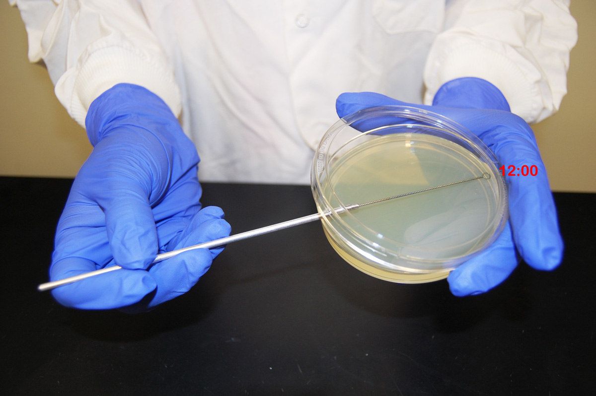
In order to avoid digging into the agar as you streak the loop over the top of the agar you must keep the loop parallel to the the agar surface. Always start streaking at the "12:00 position" (See Fig. 3C.) of the plate and streak side-to-side as you pull the loop toward you. As you follow either Fig. 4 or Fig. 5, each time you flame and cool the loop between sectors, rotate the plate counterclockwise so you are always working in the "12:00 position" of the plate. This keeps the inoculating loop parallel with the agar surface and helps prevent the loop from digging into the agar.
Fig. 3C: Streaking an Agar Plate
Keep the inoculating loop parallel with the agar and rotate the plate so you are always streaking the agar in the "12:00" position.Gary E. Kaiser, Ph.D.
Professor of Microbiology
The Community College of Baltimore County, Catonsville Campus
This work is licensed under a Creative Commons Attribution 3.0 Unported License
5. Remove the loop and immediately close the lid.
6. Resterilize the inoculating loop by placing it in the bacto-incinerator for 10 seconds. (See Fig. 3D.)
Fig. 3D: Using a Bacto-incinerator to Sterilize the Inoculating Loop
Sterilize the inoculating loop by placing it in the bacto-incinerator for 10 seconds.Gary E. Kaiser, Ph.D.
Professor of Microbiology
The Community College of Baltimore County, Catonsville Campus
This work is licensed under a Creative Commons Attribution 3.0 Unported License
Examples of plates showing isolated colonies. See Figs. 3E-3G.
Fig. 3E: Serratia marcescens on Trypticase Soy Agar; Three Sector Method Fig. 3F: Escherichia coli on Trypticase Soy Agar; Three Sector Method
Fig. 3G: Fecal Specimen on Blood Agar; Three Sector Method
Note Isolated colonies.
Note Isolated colonies.
Note Isolated colonies. Serratia marcescens on trypticase soy agar. © D. Sue Katz, author.
Licensed for use, ASM MicrobeLibrary.Escherichia coli on trypticase soy agar. © D. Sue Katz, author.
Licensed for use, ASM MicrobeLibrary.Fecal specimen on trypticase soy agar. © D. Sue Katz, author.
Licensed for use, ASM MicrobeLibrary.
In the future, every procedure in the lab will be done using similar aseptic technique.
B. FORMS OF CULTURE MEDIA
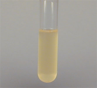
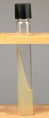
1. Broth tubes (see Fig. 4A) are tubes containing a liquid medium. A typical nutrient containing broth medium such as Trypticase Soy broth contains substrates for microbial growth such as pancreatic digest of casein, papaic digest of soybean meal, sodium chloride, and water.
Fig. 4A: Broth Culture of a Bacterium
Gary E. Kaiser, Ph.D.
Professor of Microbiology
The Community College of Baltimore County, Catonsville Campus
This work is licensed under a Creative Commons Attribution 3.0 Unported License
After incubation, growth (development of many cells from a few cells) may be observed as one or a combination of three forms:
a. Pellicle: A mass of organisms is floating on top of the broth. (See Fig. 4A.)
b. Turbidity: The organisms appear as a general cloudiness throughout the broth. (See Fig. 4B.).
c. Sediment: A mass of organisms appears as a deposit at the bottom of the tube. (See Fig. 4C.)
Fig. 4B: Broth Culture Showing Pellicle
Fig. 4C: Broth Culture Showing Turbidity
Fig. 4D: Broth Culture Showing Sediment
Gary E. Kaiser, Ph.D.
Professor of Microbiology
The Community College of Baltimore County, Catonsville Campus
This work is licensed under a Creative Commons Attribution 3.0 Unported License
Gary E. Kaiser, Ph.D.
Professor of Microbiology
The Community College of Baltimore County, Catonsville Campus
This work is licensed under a Creative Commons Attribution 3.0 Unported License
Gary E. Kaiser, Ph.D.
Professor of Microbiology
The Community College of Baltimore County, Catonsville Campus
This work is licensed under a Creative Commons Attribution 3.0 Unported License
2. Slant tubes (see Fig. 5A)are tubes containing a nutrient medium plus a solidifying agent, agar-agar. The medium has been allowed to solidify at an angle in order to get a flat inoculating surface (see Fig. 5B).
Fig. 5A: An Agar Slant Tube (Side View)
Fig. 5B: An Agar Slant Culture
Gary E. Kaiser, Ph.D.
Professor of Microbiology
The Community College of Baltimore County, Catonsville Campus
This work is licensed under a Creative Commons Attribution 3.0 Unported License
Gary E. Kaiser, Ph.D.
Professor of Microbiology
The Community College of Baltimore County, Catonsville Campus
This work is licensed under a Creative Commons Attribution 3.0 Unported License
3. Stab tubes (deeps) are tubes of hardened agar medium which are inoculated by "stabbing" the inoculum into the agar. (See Fig. 6.)
Fig. 6: Agar Stab Culture of a Bacterium
Gary E. Kaiser, Ph.D.
Professor of Microbiology
The Community College of Baltimore County, Catonsville Campus
This work is licensed under a Creative Commons Attribution 3.0 Unported License
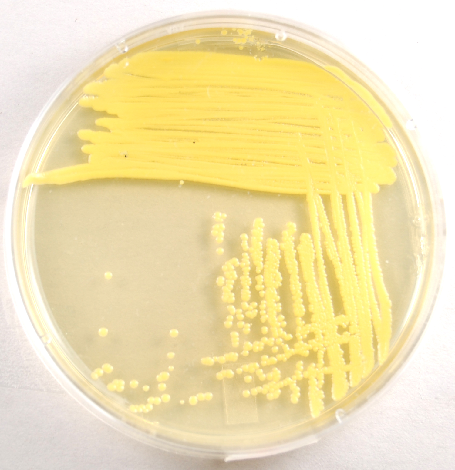
4. Agar plates (see Fig. 7) are sterile petri plates that are aseptically filled with a melted sterile agar medium and allowed to solidify. Plates are much less confining than slants and stabs and are commonly used in the culturing, separating, and counting of microorganisms. Single colonies of microorganisms on agar plates can be described using the terms found in Appendix A.
Fig. 7: Isolated Colonies of Escherichia coli growing on an agar plate
Note Isolated colonies. Escherichia coli on Luria agar. © Kevin Hedetniemi and Min-Ken Liao, authors.
Licensed for use, ASM MicrobeLibrary.
C. OXYGEN REQUIREMENTS FOR MICROBIAL GROWTH
Microorganisms show a great deal of variation in their requirements for gaseous oxygen. Most can be placed in one of the following groups:
1. Obligate aerobes are organisms that grow only in the presence of oxygen. They obtain energy from aerobic respiration.
2. Microaerophiles are organisms that require a low concentration of oxygen for growth. They obtain energy from aerobic respiration.
3. Obligate anaerobes are organisms that grow only without oxygen and, in fact, oxygen inhibits or kills them. They obtain energy from anaerobic respiration or fermentation.
4. Aerotolerant anaerobes, like obligate anaerobes, cannot use oxygen for growth but they tolerate it fairly well. They obtain energy from fermentation.
5. Facultative anaerobes are organisms that grow with or without oxygen, but generally better with oxygen. They obtain energy from aerobic respiration, anaerobic respiration, and fermentation. Most bacteria are Facultative anaerobes.
D. TEMPERATURE REQUIREMENTS
Microorganisms have a minimum and maximum temperature at which they can grow, as well as an optimum temperature where they grow best. Microorganisms can be divided into groups on the basis of their preferred range of temperature:
1. Psychrophiles are cold-loving bacteria. Their optimum growth temperature is between -5°C and 15°C. They are usually found in the Arctic and Antarctic regions and in streams fed by glaciers.
2. Mesophiles are bacteria that grow best at moderate temperatures. Their optimum growth temperature is between 25°C and 45°C. Most bacteria are mesophilic and include common soil bacteria and bacteria that live in and on the body.
3. Thermophiles are heat-loving bacteria. Their optimum growth temperature is between 45°C and 70°C and are commonly found in hot springs and in compost heaps.
4. Hyperthermophiles are bacteria that grow at very high temperatures. Their optimum growth temperature is between 70°C and 110°C. They are usually members of the Archae and are found growing near hydrothermal vents at great depths in the ocean.
E. COLONY MORPHOLOGY AND PIGMENTATION
A colony (see Fig. 8A) is a visible mass of microorganisms growing on an agar surface and usually originating from a single organism or arrangement of organisms. Different microorganisms will frequently produce colonies which differ in their morphological appearance (form, elevation, margin, surface, optical characteristics, and pigmentation). Single colonies can be described using standard terms, as listed in Appendix A.
Fig. 8A: Isolated Colonies of Escherichia coli
Fig. 8B: A Microcolony of Escherichia coli
Note the layers of the bacillus-shaped E. coli. Escherichia coli on Luria agar. © Kevin Hedetniemi and Min-Ken Liao, authors.
Licensed for use, ASM MicrobeLibrary.Escherichia coli Microcolony. © James Shapiro and Clara Hsu, authors. Licensed for use, ASM MicrobeLibrary.
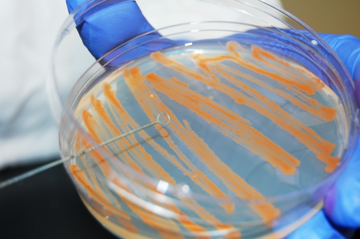
Probably the most visual characteristic is pigmentation (color). Some microorganisms produce pigment during growth and are said to be chromogenic. Often, however, formation of pigment depends on environmental factors such as temperature, nutrients, pH and moisture. For example, Serratia marcescens produces a deep red pigment at 25°C, but does not produce pigment at 37°C.
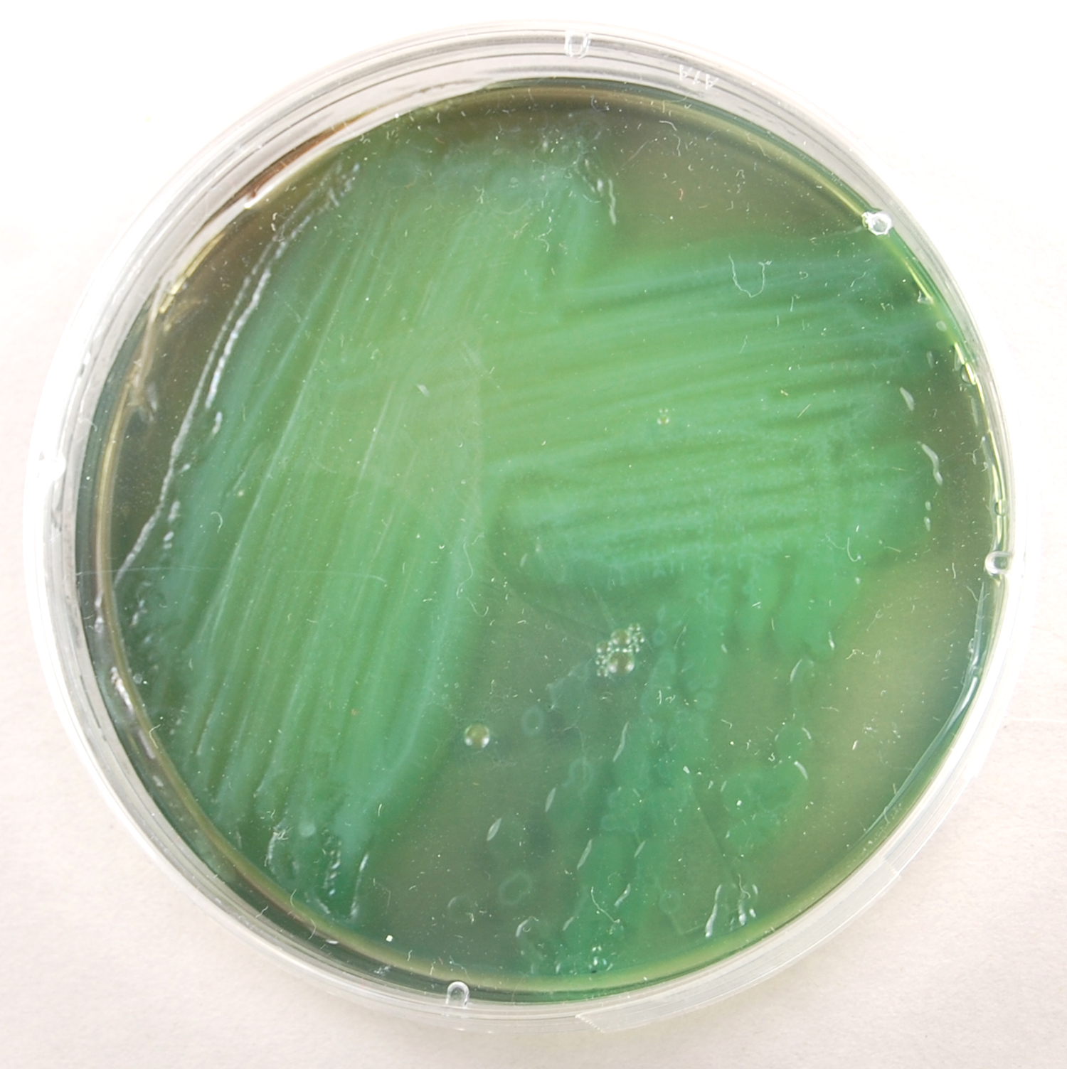
Pigments can be divided into two basic types: water-insoluble and water-soluble. If the pigment is water-insoluble (see Figs. 9A-9E), as is the case with most chromogenic bacteria, it does not diffuse out of the organism. As a result, the colonies are pigmented but the agar remains the normal color. If the pigment is water-soluble as in the case of Pseudomonas aeruginosa (see Fig. 9F and Fig. 9G), it will diffuse out of the organism into the surrounding medium causing the agar to appear pigmented. Note that pigment can generally be detected only on a medium that was originally colorless.
Fig. 9A: TSA Plate Culture of the Chromogenic Bacterium Serratia marcescens
Fig.9B: TSA Plate Culture of the Chromogenic Bacterium Micrococcus luteus
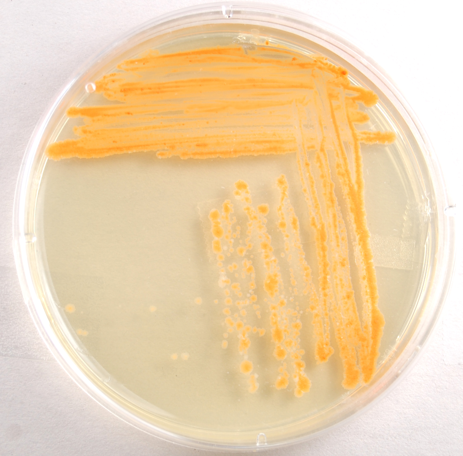
Fig. 9C: TSA Pate Culture of the Chromogenic Bacterium Mycobacterium phlei
Note red, water insoluble pigment.
Note yellow, water insoluble pigment.
Note orange, water insoluble pigment. Gary E. Kaiser, Ph.D.
Professor of Microbiology
The Community College of Baltimore County, Catonsville Campus
This work is licensed under a Creative Commons Attribution 3.0 Unported License
Gary E. Kaiser, Ph.D.
Professor of Microbiology
The Community College of Baltimore County, Catonsville Campus
This work is licensed under a Creative Commons Attribution 3.0 Unported License
Gary E. Kaiser, Ph.D.
Professor of Microbiology
The Community College of Baltimore County, Catonsville Campus
This work is licensed under a Creative Commons Attribution 3.0 Unported License
Fig. 9D: TSA Plate Culture of the Chromogenic Bacterium Micrococcus roseus
Fig. 9E: TSA Plate Culture of the Chromogenic Bacterium Staphylococcus aureus
Note pink rose, water insoluble pigment.
Note gold, water insoluble pigment. Gary E. Kaiser, Ph.D.
Professor of Microbiology
The Community College of Baltimore County, Catonsville Campus
This work is licensed under a Creative Commons Attribution 3.0 Unported License
Last updated: August, 2017
Please send comments and inquiries to Dr. Gary KaiserGary E. Kaiser, Ph.D.
Professor of Microbiology
The Community College of Baltimore County, Catonsville Campus
This work is licensed under a Creative Commons Attribution 3.0 Unported License
Last updated: August, 2017
Please send comments and inquiries to Dr. Gary Kaiser
Fig. 9F: TSA Plate Culture of the Chromogenic Bacterium
Pseudomonas aeruginosaFig. 9G: Pseudomonas aeruginosa Growing on Trypticase Soy Agar
Note green, water soluble pigment.
Note green, water soluble pigment. Gary E. Kaiser, Ph.D.
Professor of Microbiology
The Community College of Baltimore County, Catonsville Campus
This work is licensed under a Creative Commons Attribution 3.0 Unported License
Gary E. Kaiser, Ph.D.
Professor of Microbiology
The Community College of Baltimore County, Catonsville Campus
This work is licensed under a Creative Commons Attribution 3.0 Unported License
MEDIA
Trypticase Soy Broth tubes (4), Trypticase Soy Agar slant tubes (4), Trypticase Soy Agar stab tubes (4), and Trypticase Soy Agar plates (7).
ORGANISMS
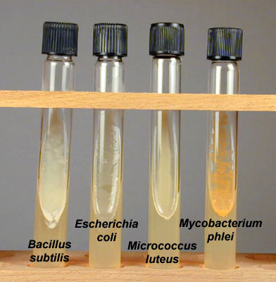
Trypticase Soy Broth cultures of Bacillus subtilis, Escherichia coli and Micrococcus luteus, and Trypticase Soy Agar plate cultures of Mycobacterium phlei.
PROCEDURE (to be done in pairs)
Video review - Procedure for Today's Lab
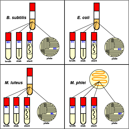
1. Aseptically inoculate one Trypticase Soy Broth tube, one Trypticase Soy Agar slant tube, one Trypticase Soy Agar stab tube, and one Trypticase Soy Agar plate with B. subtilis. (See Fig. 10.)
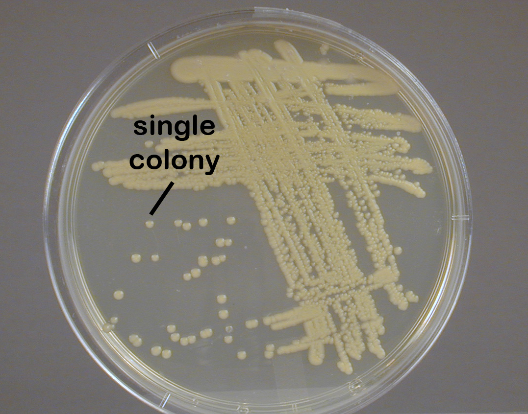
Remember to label all tubes with a wax marker. When streaking the agar plates, use either of the patterns shown above in Figs. 4A-4C or Figs. 5A-5E. This procedure is termed streaking for isolation and has a diluting effect. The friction of the loop against the agar causes organisms to fall off the loop. Near the end of the streaking pattern, individual organisms become separated far enough apart on the agar surface to give rise to isolated single colonies after incubation.
2. Aseptically inoculate one Trypticase Soy Broth tube, one Trypticase Soy Agar slant tube, one Trypticase Soy Agar stab tube, and one Trypticase Soy Agar plate with E. coli. (See Fig. 10)
3. Aseptically inoculate one Trypticase Soy Broth tube, one Trypticase Soy Agar slant tube, one Trypticase Soy Agar stab tube, and one Trypticase Soy Agar plate with M. luteus. (See Fig. 10.)
4. Aseptically inoculate one Trypticase Soy Broth tube, one Trypticase Soy Agar slant tube, one Trypticase Soy Agar stab tube, and one Trypticase Soy Agar plate with M. phlei. (See Fig. 10.)
Fig. 10: Procedure for Lab 2
Inoculate a separate sterile broth tube, stab tube, slant tube, and petri plate with each of the 4 bacteria we are using today as shown in the illustration above.Gary E. Kaiser, Ph.D.
Professor of Microbiology
The Community College of Baltimore County, Catonsville Campus
This work is licensed under a Creative Commons Attribution 3.0 Unported License
5. Incubate all the tubes and plates inoculated with B. subtilis, E. coli, M. luteus, and M. phlei at 37°C. Place the tubes in your dedicated test tube rack. Incubate the petri plates upside down (lid on the bottom) and stacked in the petri plate holder on the shelf of the 37°C incubator corresponding to your lab section. Incubating the plates upside down prevents condensing water from falling on the growing colonies and causing them to run together. (Store your test tube rack on your incubator shelf when not in use.)
6. In order to illustrate that microorganisms are all around us and to demonstrate the necessity for proper aseptic technique, contaminate three Trypticase Soy Agar plates as follows:
a. Remove the lid from the first agar plate and place the exposed agar portion in or out of the building for the duration of today's lab. Replace the lid, label the plate "air", and incubate it upside-down at room temperature. Do this plate first. A typical "air" plate is shown in Fig. 12A.
b. Using a wax marker, divide a second petri plate in half. You and your partner both moisten a sterile cotton swab in sterile water. Rub your swab over some surface in the building or on yourself. Use this swab to inoculate your half of the second agar plate. Label the plate and incubate upside-down at room temperature. A plate showing growth from a swab of the bottom of a shoe is shown in Fig. 12B.
c. With a wax marker, divide a third petri plate into quarters and label as shown in Fig. 12C. On your half of the plate, first rub the fingers of one of your gloved hands over your "glove" quadrant. Remove that glove and rub your fingers over your "fingers" quadrant. Your partner will do the same on his or her half of the plate. Label the plate and incubate upside-down in your petri plate holder at 37°C. Do this plate last.
Fig. 12A: Microorganisms growing on an agar plate exposed to the air for one hour.
Fig. 12B: An agar plate showing microorganisms growing from the bottom of a shoe.
Fig. 12C: Inoculation of Your "Fingers" Plate.
Gary E. Kaiser, Ph.D.
Professor of Microbiology
The Community College of Baltimore County, Catonsville Campus
This work is licensed under a Creative Commons Attribution 3.0 Unported License
Gary E. Kaiser, Ph.D.
Professor of Microbiology
The Community College of Baltimore County, Catonsville Campus
This work is licensed under a Creative Commons Attribution 3.0 Unported License
Gary E. Kaiser, Ph.D.
Professor of Microbiology
The Community College of Baltimore County, Catonsville Campus
This work is licensed under a Creative Commons Attribution 3.0 Unported License
RESULTS
1. Draw and describe the growth seen in each of the four broth cultures.
Growth =
Growth =
Growth =
Growth =
2. Observe the growth in the slant cultures and stab cultures for pigmentation and purity.
Agar Slant Cultures of Bacteria Gary E. Kaiser, Ph.D.
Professor of Microbiology
The Community College of Baltimore County, Catonsville Campus
This work is licensed under a Creative Commons Attribution 3.0 Unported License
3. Using the terms in the Appendix A, compare a single colony of each of the four bacteria used in today's lab.
Single Colonies of Bacillus subtilis
Isolated Colonies of Escherichia coli
Single Colonies of Micrococcus luteus
Single Colonies of Mycobacterium phlei
Gary E. Kaiser, Ph.D.
Professor of Microbiology
The Community College of Baltimore County, Catonsville Campus
This work is licensed under a Creative Commons Attribution 3.0 Unported License
Image: Escherichia coli on Luria agar. © Kevin Hedetniemi and Min-Ken Liao, authors.
Licensed for use, ASM MicrobeLibrary.Gary E. Kaiser, Ph.D.
Professor of Microbiology
The Community College of Baltimore County, Catonsville Campus
This work is licensed under a Creative Commons Attribution 3.0 Unported License
Gary E. Kaiser, Ph.D.
Professor of Microbiology
The Community College of Baltimore County, Catonsville Campus
This work is licensed under a Creative Commons Attribution 3.0 Unported License
characteristics Form of colony Elevation Margin (edge) Surface Optical characteristics Pigmentation
4. Observe the results of the three "contamination" plates and note the differences in colony appearances.
5. Observe the demonstration plates of chromogenic bacteria and state the color and water-solubility of each pigment.
organism Micrococcus luteus Micrococcus roseus Mycobacterium phlei Serratia marcescens Staphylococcus aureus Pseudomonas aeruginosa Fig. 10B and Pseudomonas aeruginosa Fig. 10C
PERFORMANCE OBJECTIVES FOR LAB 2
After completing this lab, the student will be able to perform the following objectives:
DISCUSSION
1. Define the following terms: pure culture, sterile medium, inoculum, aseptic technique, and colony.
2. State and define the three types of growth that may be seen in a broth culture.
3. Define the following terms: obligate aerobe, microaerophile, obligate anaerobe, aerotolerant anaerobe, and Facultative anaerobe.
4. Define the following terms: psychrophile, mesophile, thermophile, and hyperthermophile.
5. Define the following terms: chromogenic, water-soluble pigment, and water-insoluble pigment.
PROCEDURE
1. Using an inoculating loop, demonstrate how to aseptically remove some inoculum from either a broth tube, slant tube, stab tube, or petri plate, and inoculate a sterile broth tube, slant tube, stab tube, or petri plate without introducing outside contamination.
2. Label all tubes and plates and place them on the shelf in the incubator corresponding to your lab section.
3. Return all class pure cultures to the instructor’s lab bench.
4. Dispose of all materials when the experiment is completed, being sure to remove all markings from the glassware. Place all culture tubes in the plastic baskets in the biohazard hood and all petri plates in the buckets in the biohazard hood.
RESULTS
1. Recognize and identify the following types of growth in a broth culture: pellicle, turbidity, sediment, and any combination of these.
2. State the color and water-solubility of pigment seen on a plate culture of a chromogenic bacterium.
SELF-QUIZ
Microbiology Laboratory Manual by Gary E. Kaiser, PhD, Professor of Microbiology
is licensed under a Creative Commons Attribution 4.0 International License.
Last updated: March, 2023
Please send comments and inquiries to Dr. Gary Kaiser